
At Southern Eye Group, protecting your vision starts with seeing the full picture. That’s why we offer optomap Ultra-Widefield (UWF™) Retinal Imaging, an advanced diagnostic technology that provides a broader, more detailed view of the retina—often without traditional dilation.
Serving patients in Mobile, Alabama and throughout the Gulf Coast, Southern Eye Group uses optomap imaging to support early detection, accurate documentation, and long-term monitoring of eye health, all while providing a more comfortable and efficient exam experience.
- What Is optomap Retinal Imaging?
- How optomap Works
- What Conditions Can optomap Help Identify?
- Why Annual Eye Exams Are So Important
- Benefits of optomap Retinal Imaging
- optomap vs. Traditional Dilated Eye Exams
- Why Southern Eye Group Uses optomap Technology
- Frequently Asked Questions About optomap Retinal Imaging
What Is optomap Retinal Imaging?
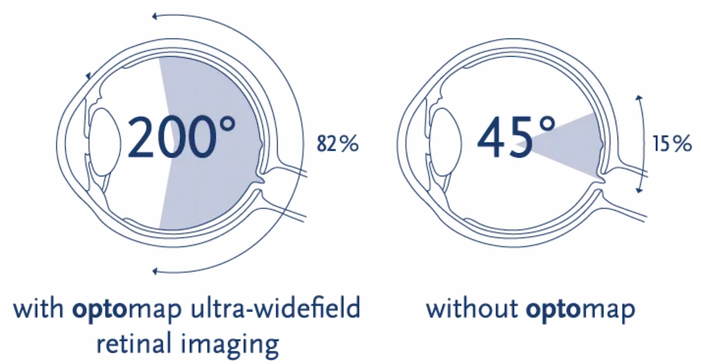
Optomap is a digital retinal imaging system that captures a high-resolution panoramic image of the retina in a single scan. Unlike traditional retinal exams or standard retinal photography, which typically show only the central portion of the retina, optomap can image more than 80% (up to 200 degrees) of the retina at
 Trusted Source
Optomap Physician Information
Go to Source
once.
Trusted Source
Optomap Physician Information
Go to Source
once.
This expanded view allows eye care providers to evaluate both the central and peripheral retina—areas where certain eye conditions may first develop. The images are available immediately and securely stored for future comparison.
How optomap Works
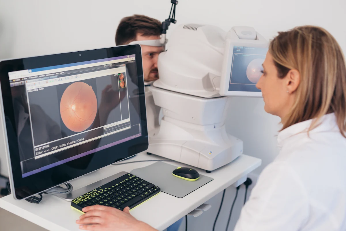
Using advanced scanning laser technology, optomap captures a detailed retinal image in just seconds. During the scan, you’ll briefly look into the imaging device while the system records a wide view of your retina.
In many cases, the scan can be completed without dilation drops, helping reduce discomfort, light sensitivity, and overall appointment time. Your provider can review the images with you during your visit, making it easier to understand your eye health.
What Conditions Can optomap Help Identify?
Because optomap captures a wide view of the retina—including the peripheral areas—it can assist in identifying signs of various eye conditions that may not cause early symptoms, such as:
- Diabetic retinopathy
- Macular degeneration
- Retinal tears or detachments
- Glaucoma-related changes
- Retinal vascular abnormalities
- Peripheral retinal lesions
- Early signs of systemic conditions that affect the eyes
While optomap does not diagnose conditions on its own, it provides valuable visual information that helps your eye care provider evaluate retinal health and determine whether further testing or treatment is needed.
Why Annual Eye Exams Are So Important
Many eye diseases develop gradually and may not cause noticeable symptoms in their early stages. By the time vision changes occur, damage may already be present.
Annual comprehensive eye exams allow your provider to:
- Detect subtle changes in eye health early
- Monitor existing conditions over time
- Establish a baseline for future comparison
- Evaluate how systemic health issues may be affecting your eyes
Optomap imaging enhances yearly exams by offering a detailed, documented view of the retina, helping your provider track changes from one visit to the next and make informed care recommendations.
Benefits of optomap Retinal Imaging
A Broader View of Retinal Health
The optomap produces an image that is unique and provides Southern Eye Group with a high-resolution 200° image in order to ascertain the health of your retina. This is much wider than a traditional 45° image.
Comfortable Alternative to Traditional Dilation
For many patients, optomap can be performed without dilation, meaning:
- No stinging drops
- No prolonged light sensitivity
- Less disruption to daily activities
Efficient, Patient-Friendly Experience
The scan takes only seconds per eye, which may help shorten overall appointment time.
Long-Term Monitoring
Optomap images are stored as part of your medical record, allowing your Southern Eye Group provider to compare images over time and monitor retinal changes.
Suitable for Most Patients
Optomap imaging is useful for a wide range of patients—not just those with known retinal conditions—and is often included as part of routine eye care.
optomap vs. Traditional Dilated Eye Exams
While optomap offers many advantages in comfort and efficiency, it does not replace a comprehensive dilated eye exam in all cases. Certain conditions still require dilation for the most thorough evaluation. Your provider will recommend the appropriate exam approach based on your individual eye health and risk factors.

Why Southern Eye Group Uses optomap Technology
Southern Eye Group is committed to delivering high-quality, technology-driven eye care across the region. With multiple locations, we proudly serve patients in Mobile, Alabama and throughout the Gulf Coast, offering access to advanced diagnostic tools close to home.
Our use of optomap reflects our commitment to:
- Early identification of potential eye concerns
- Thorough and efficient evaluations
- Patient comfort and education
- Continuity of care year after year
Frequently Asked Questions About Optomap Retinal Imaging
What does optomap retinal imaging show?
Optomap captures a wide, high-resolution image of the retina, including peripheral areas that may not be visible with traditional imaging, allowing for a more comprehensive evaluation.
Is optomap an alternative to eye dilation or a replacement for a dilated eye exam?
In many cases, Optomap retinal imaging can be performed without dilation, offering a more comfortable and convenient experience. However, Optomap does not replace a comprehensive dilated eye exam in all situations. Your provider may still recommend dilation based on your eye health, symptoms, or clinical findings.
Who should have optomap retinal imaging?
Optomap is beneficial for most patients, including those with diabetes, high blood pressure, vision changes, or a family history of eye disease, as well as patients seeking thorough routine eye care.
How often should I have retinal imaging?
Many patients receive retinal imaging as part of their annual eye exam. Your provider will recommend the appropriate frequency based on your eye health and risk factors.
Are Optomap images saved for future visits?
Yes. Images are stored securely and can be compared over time to monitor changes in retinal health.
Is the optomap included in my visit?
The optomap is not covered by insurance and is provided for a fee of $39.00.

Contact Southern Eye Group
To learn more about optomap Ultra-Widefield Retinal Imaging or to schedule your annual eye exam, contact Southern Eye Group today.
Serving patients in Mobile, Alabama and throughout the Gulf Coast, our team is here to support your vision at every stage of life.
1 Optomap Physician Information. Available: https://www.optos.com/products/. Accessed January 3, 2026.
The doctors at Southern Eye Group have either authored or reviewed and approved this content.
Page Updated: